
A Lump Was Found on My Kidney Scan: What Incidental Kidney Masses and Cysts Really Mean?
An ultrasound or CT scan performed for an unrelated concern, such as abdominal pain or a gallbladder problem, may unexpectedly identify a “lesion”, “cyst” or “mass” in the kidney. Such findings can cause concern, but they do not always indicate cancer. Some are benign, while others require further imaging or specialist assessment. This blog explains how incidental kidney findings are evaluated and what the next steps may involve.
What Is an Incidental Kidney Finding?
An “incidental” finding is discovered by chance on a scan performed for another reason. As ultrasound, CT and MRI scans have become more widely used, kidney cysts and small masses are detected more often. Some kidney cancers are discovered before symptoms develop. Detecting a lesion at an earlier stage can broaden management options, although its significance depends on the type and characteristics of the finding.
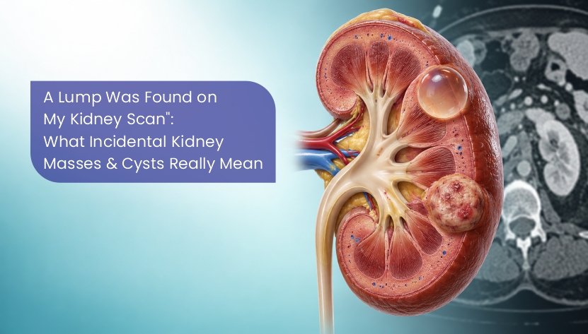
Incidental kidney findings fall into two broad groups: cysts, which are fluid-filled sacs, and solid masses, which are made of tissue.
Kidney Cysts: Understanding Simple and Complex Cysts
Simple kidney cysts are common and become more frequent with age. A simple cyst is typically a thin-walled sac containing fluid. When imaging confirms that a cyst is simple, it is considered benign and usually needs no treatment or follow-up. Occasionally, a large cyst causes symptoms and may require assessment or intervention.
Some cysts, however, look more complicated on a scan. They may have thick walls, internal divisions (septa), calcium deposits or solid areas. These are called complex cysts, and they need closer evaluation.
The Bosniak Classification: How Cysts Are Graded
Urologists and radiologists use the Bosniak classification to describe kidney cysts, based on how they look on a contrast CT or MRI scan. It helps estimate the risk of cancer and guides management:
- Bosniak I: simple cyst with a thin wall and clear fluid; benign, no follow-up needed
- Bosniak II: minimally complex, with a few thin septa or fine calcification; benign, usually no follow-up needed
- Bosniak IIF: slightly more complex ("F" stands for follow-up); low risk, monitored with periodic scans
- Bosniak III: thickened or irregular enhancing walls or septa; a meaningful risk of malignancy, so surveillance or intervention may be discussed depending on the lesion and patient
- Bosniak IV: enhancing nodular or solid components; a high likelihood of malignancy, requiring specialist discussion of management options
This grading is why doctors sometimes request a further scan before making any recommendation. An ultrasound alone often cannot fully characterise a complex cyst.
Solid Kidney Masses: What Could They Be?
A solid mass needs more careful attention than a cyst, but it is not automatically cancer. Possible causes include:
- Renal cell carcinoma (kidney cancer): the most common solid kidney tumour in adults, though many small ones are low-grade and slow-growing
- Angiomyolipoma: a benign tumour made of fat, blood vessels and muscle, usually recognised on CT by its fat content
- Oncocytoma: a benign tumour that can be difficult to tell apart from cancer on imaging
- Other causes: infections, abscesses, or rarely spread from cancers elsewhere
Research on surgically removed small solid kidney masses shows that some are benign. Some malignant small renal masses grow slowly, but growth patterns differ and cannot reliably establish whether a lesion is harmless. Imaging findings and individual risk factors guide further evaluation.
What Tests Might You Need?
Depending on what the first scan shows, your urologist may recommend:
- A dedicated contrast CT or MRI of the kidneys: to see whether the lesion takes up contrast (enhances), which is a key sign of solid tissue
- Contrast-enhanced ultrasound: helpful for some complex cysts and for patients who cannot have CT contrast
- Blood and urine tests: to check kidney function and look for blood in the urine
- Renal mass biopsy: in selected cases, a small needle sample can help decide between surveillance and treatment
Treatment Options
Management is tailored to the size and appearance of the lesion, as well as the patient's age, kidney function and overall health:
- Active surveillance: regular scans to monitor small masses, particularly in older patients or those with other health problems
- Partial nephrectomy: removal of only the tumour while preserving the rest of the kidney, often performed with robotic or laparoscopic surgery
- Thermal ablation: freezing (cryoablation) or heating (radiofrequency or microwave ablation) the tumour through a needle, for selected small tumours
- Radical nephrectomy: removal of the whole kidney, generally reserved for larger or more complex tumours
Where clinically appropriate, preserving healthy kidney tissue may help maintain long-term kidney function. The safest approach depends on tumour characteristics and the patient’s overall condition.
Understanding the Prognosis of Kidney Masses
For some small kidney cancers confined to the kidney, treatment can be associated with favourable long-term outcomes. Prognosis still depends on the tumour type, stage, grade, kidney function and overall health. A mass discovered before symptoms appear may offer different management options, but an incidental finding does not automatically mean the disease is benign or curable.
Questions to Ask Your Urologist
- Is this a simple cyst, a complex cyst or a solid mass?
- What is its size and, for cysts, its Bosniak category?
- Do I need further imaging or a biopsy?
- Is monitoring a safe option for me?
- If treatment is needed, can my kidney be preserved?
Conclusion
A kidney cyst or mass discovered unexpectedly on a scan can represent several different conditions. Simple cysts are usually benign, but complex cysts and solid masses may need further investigation, monitoring or treatment. An appropriate next step is to have the report and images reviewed by a qualified clinician, who can advise on any further tests and individual management options.
Contact Us
The Gujarat Uro Oncology Associates team offers expert consultation, diagnosis, second opinions, and treatment planning for urological cancers. To learn more or book an appointment, contact 91 75730 04253 or email bakshihemang@yahoo.co.in.